
Kidney Stone Diagnosis: Complete Guide to Sonography and Imaging Tests
When you experience sharp pain in your lower back or side, blood in your urine, or difficulty passing urine, your doctor might suspect kidney stones. But how do doctors actually see these tiny stones inside your body? At Arka Anugraha Hospital in Bangalore, we use advanced imaging tests to accurately diagnose kidney stones and create the best treatment plan for you.
Understanding kidney stone sonography and other imaging methods can help you feel more prepared and less anxious about your diagnosis.
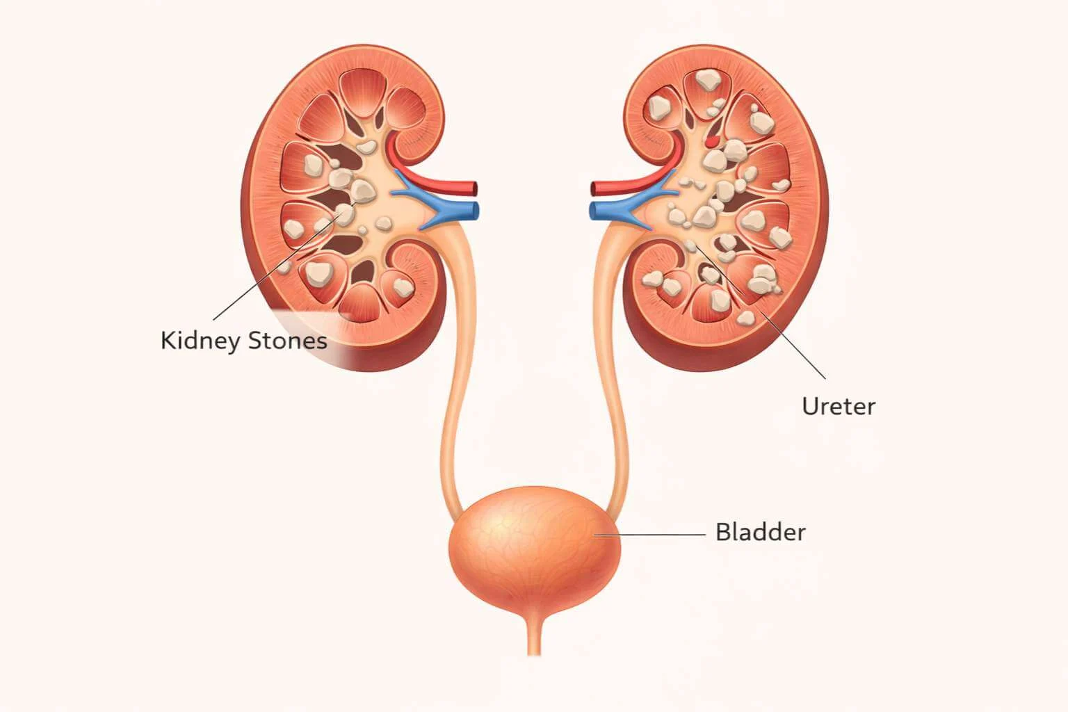
What Is Kidney Stone Sonography (Ultrasound)?
Kidney stone sonography is a safe and painless way to look inside your body using sound waves. Unlike X-rays, it does not use radiation, making it completely safe for pregnant women and children.
How It Works
A technician applies gel to your abdomen and moves a transducer across your skin. Sound waves bounce off stones, creating images on a computer. It takes about 15-30 minutes and is painless.
Comparing Imaging Tests
Different situations call for different tests. Here is a breakdown of the three main methods used at Arka Anugraha Hospital:
Ultrasound (Sonography)
Pros: No radiation, safe for pregnancy/children, detects blockages.
Cons: Can miss small stones (<4mm) or stones in the ureter.
X-Ray (KUB)
Pros: Quick, low cost, minimal radiation.
Cons: Misses uric acid stones and small stones. Good for tracking calcium stones.
CT Scan
Pros: Most accurate (>95%), finds tiny stones (1mm), shows exact location.
Cons: Higher radiation (though modern low-dose protocols are safe), higher cost.
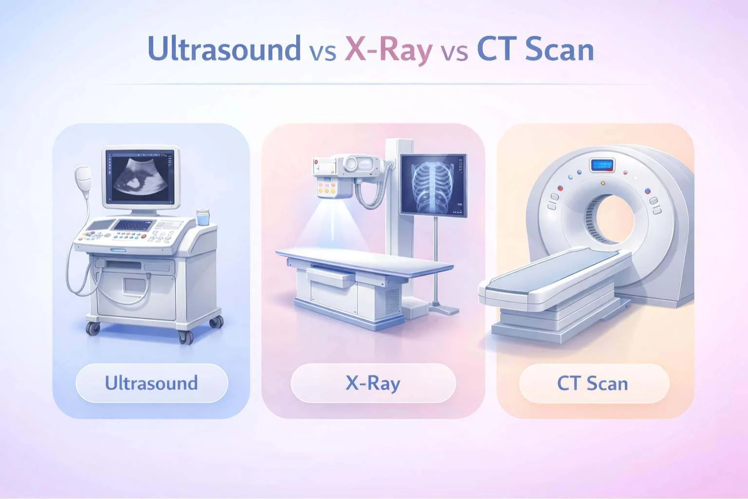
Which Test is Right for You?
Understanding Your Results
After imaging, a radiologist analyzes the pictures. Key factors include:
- Size: Stones <5mm often pass naturally. >10mm usually need surgery.
- Location: Stones in the ureter cause pain; stones in the kidney may be silent.
- Density: Dense stones (on CT) are likely calcium; less dense may be uric acid.
How to Prepare for Your Test
Preparation Guidelines
- Ultrasound: Drink plenty of water beforehand. A full bladder helps create clearer images.
- X-Ray: No special prep. Remove metal jewelry.
- CT Scan: Follow doctor's instructions. You may need to fast for a few hours if contrast dye is used. Inform them of any allergies.
How Imaging Guides Treatment
Watchful Waiting: If images show small stones without blockage, fluids and pain meds are prescribed.
Surgery: Large stones or blockages seen on CT scans may require procedures like shock wave therapy or ureteroscopy.
Why Ongoing Imaging Matters
If you've had stones before, you're at higher risk. Regular ultrasounds (every 6-12 months) can catch new stones early when they are easier to treat.
Expert Diagnosis at Arka Anugraha Hospital
Accurate imaging is the foundation of effective treatment. Our advanced technology and experienced team ensure you get the best care.
If you have symptoms or questions about kidney stones, contact us today.
Book Your Scan Now